
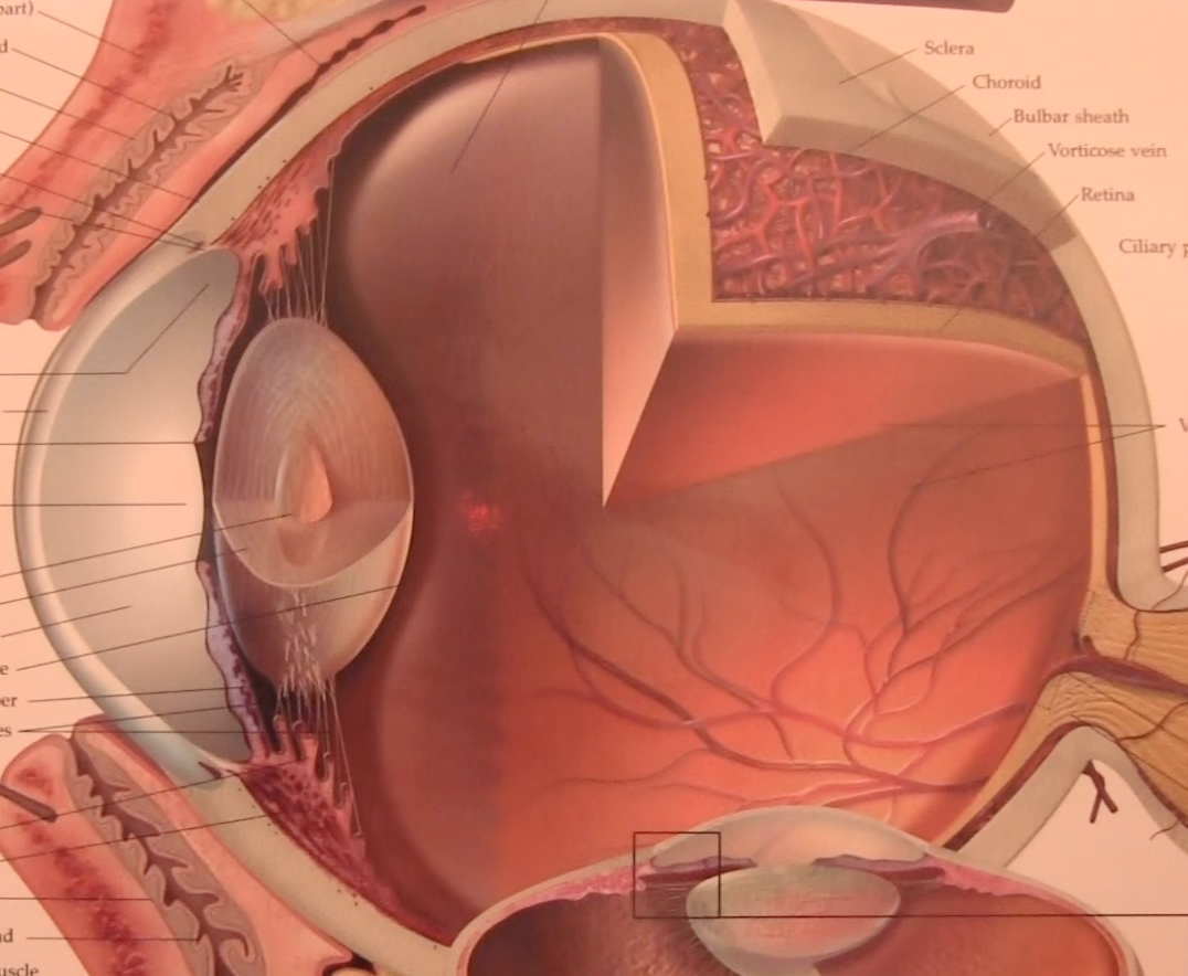
Visual health consists of many aspects of the eye, including parts you cannot see on the surface of the eye. Learning the structures and functions of the eye are the best way to understand how to keep the eyes healthy and why eye diseases may occur. Here is an overview of the anatomy of the human eye.
Eyelid
The eyelid consists of two movable folds which protect the eye. At the edge of the eyelid are 2 to 3 rows of eyelashes, called CILIA. The eyelids consist of multiple layers. From outer to inner most layer: the skin, subcutaneous areolar, orbicularis layer, orbital septum, superior levator palpebrae muscle, muscle of muller, tarsal plate, and palpebral conjunctiva.
Fun fact: When you see someone flipping their eyelid over, that pinkish area is called the palpebral conjunctiva. This is an immunologically active layer filled with macrophages and mast cells, which provide a defense against foreign invaders.
Lacrimal System
The lacrimal system produces and drains the tears. The LACRIMAL GLAND (located behind the eyebrow) is divided into the orbital and palpebral portion and secretes the tears. The nasolacrimal drainage system is located towards the nose and is what drains the tears. The little hole at the edge of the eyelid is called the PUNCTA. This drains into the canaliculi, into the lacrimal sac, and down the lacrimal duct. The CARUNCLE is the pink part on the corner of your eye on the nose side. There’s little hair follicles there and where the debris and gunk collect in the morning.
Fun fact: Infants can sometimes get a nasolacrimal duct obstruction, which prevents the tears from draining through this system and causes yellowish discharge and tearing. This may spontaneously resolve, or will require treatment at the Optometrist’s office.
Extraocular Muscles
The eye muscles attach to the SCLERA, which is a white fibrous protective outer layer of the eye. The muscles include: superior rectus (top), inferior rectus (bottom), medial rectus (nose side), lateral rectus (ear side), superior oblique (top), and inferior oblique (bottom). These 6 muscles are how you are able to move your eyes up, down, left, right, turn inwards and turn outwards. All the recti muscles originate at the COMMON TENDINOUS RING.
Cornea
The cornea is the transparent and front part of the eye. There is a tear film resting on top. The cornea consists of multiple layers, including the epithelium, basement membrane, bowman’s layer, stroma, decemet’s membrane, and endothelium. Each layer has a different structure and function.
Fun fact: This is the layer in which LASIK and PRK are performed for refractive surgery. The cornea also has a much greater nerve supply than the skin. A cut on the front surface of the eye, called a corneal abrasion, will cause severe pain due to all the nocioceptors which innervate the cornea.
Conjunctiva
The conjunctiva is a thin, translucent membrane over the sclera.
Iris
The iris is the colored part of your eye. Iris color is determined by the amount of melanin in melanocytes. Greater amounts of melanin give a browner appearance and less amounts of melanin can give a blue or green appearance. Hazel eyes are in between. The hole in the center (what you see as a black circle) is the PUPIL. Between the cornea and iris is the ANTERIOR CHAMBER and angle. This is the site for aqueous filtration in the eye and one of the areas affected in glaucoma.
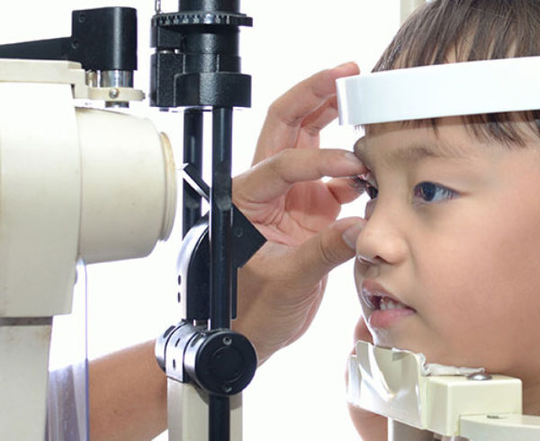
Fun fact: During an eye exam, eye drops will be used to dilate the pupil, making it larger, thus making the posterior structures of the eye visible to detect any eye diseases.
Choroid
The choroid is one of the outer layers of the eye. It consists of multiple layers and has a large blood supply. Diseases can occur here, including melanomas, age-related macular degeneration and histoplasmosis.
Ciliary Body
The ciliary body produces aqueous humor, a fluid that nourishes parts of the eye. It includes the PARS PLICATA and PARS PLANA, which produces fibers that connect to the crystalline lens.
Crystalline Lens
The crystalline lens is a transparent avascular structure. It is one of the important structures for good vision. It has the ability to accommodate, which means changing focus of your eyes on objects at a distance and then on objects up close and vice versa. When you are young, the lens is transparent and has the ability to accommodate well. At around 40 years of age, presbyopia develops due to an aging process that occurs on this structure. The lens loses the ability to accommodate, making things up close blurry and requiring reading glasses to see near.
Fun Fact: This is the structure on which cataracts occur. Cataracts develop from sun damage, systemic disease, and older age, causing the lens to change color and lose its transparency, thus, making it difficult to see.
Vitreous
The vitreous is the largest structure of the eye. It’s main components are hyaluronic acid and collagen. With age, the collagen clumps up, causing floaters, which can be seen as small objects floating in your vision. When the posterior vitreous pulls away from the retina, a posterior vitreous detachment can occur and an increased amount of floaters may be seen.
Retina
The retina is one of the posterior structures of the eye. Light energy goes through the eye, is absorbed in the retina, and transformed into chemical energy. The retina consists of photoreceptors, bipolar, and ganglion cells. There are multiple layers in the retina. The photoreceptor layer consists of RODS, which provides night vision and peripheral vision, and CONES, which provides color vision and central vision. There is also a blood supply in the retina. The branches of the central retinal artery supply the inner nuclear layer and the nerve fiber layer.
Fun fact: In uncontrolled diabetes, hemorrhages, which are blood vessel breaks and leakages, can occur in the retina and lead to decreased vision. This is why it is important to have your eyes examined at least once a year if you have diabetes to prevent vision loss. Retinal detachments can also occur at the retina, causing symptoms such as flashes of light. This is a serious eye condition which warrants treatment.
Macula
The macula is a dark, pigmented region which allows you to see color and detail. It consists of zeaxanthin and lutein pigments.
Fun fact: This area is affected in age-related macular degeneration, which will initially be treated with antioxidants and zinc therapy.
Fovea
In the center of the macula, there is a yellowish dot, called the fovea. This area has the highest concentration of cones, which is responsible for your best and sharpest central vision.
Fun fact: When you are reading or looking at an object, you are using your fovea to see the words and images clearly.
Optic Nerve
The optic nerve is commonly known as your “blind spot.” It consists of axons of ganglion cells which course all the way to your brain. This is how visual information is brought from the eye to the brain.
Fun fact: This structure is affected in glaucoma, which causes damage to the optic nerve and can lead to peripheral vision loss.
Blood supply
Arterial system:
There is a blood supply from the heart to the eye and back to the heart. The COMMON CAROTID ARTERY branches into the internal and external carotid artery and supplies blood to areas of the neck, head, and face. The INTERNAL CAROTID ARTERY becomes the OPHTHALMIC ARTERY and supplies the eye. The CENTRAL RETINAL ARTERY goes through the optic nerve and supplies the inner retina (inner nuclear layer and nerve fiber layer). The SHORT POSTERIOR ARTERIES supply the posterior parts of the eye (choroid and macula). The LONG POSTERIOR ARTERIES supply the anterior parts of the eye (iris, ciliary body, and anterior part of the choroid).
Venous system:
The venous system drains the eye and brings blood back to the heart. The CENTRAL RETINAL VEIN drains the central retinal artery out of the optic nerve to the cavernous sinus. The VORTEX VEINS are located in each quadrant posterior to the equator and drain the choroid.
Fun fact: Systemic diseases which affect the blood vessels of the heart can also affect blood vessels in the eye. Diabetes, hyptertension, and high cholesterol can all have ocular manifestations. It is important to visit your optometrist to get a comprehensive eye exam, dilate your eyes, and check the health of your eyes.
Bones of the Orbit
The bones of the orbit protect the eye. The roof, or top of the orbit, includes the frontal bone and lesser wing of the sphenoid bone. The lateral wall consists of the greater wing of sphenoid and the zygomatic bone. The floor, or bottom consists of the maxillary, palatine, and zygomatic bones. The medial wall consists of the ethmoid, lacrimal, maxilla, and sphenoid bones.
Fun fact: The floor is where fractures most likely occur because it is the weakest bone in the orbit. This is often fractured after getting in a fight or punched in the eye.
Nerve supply
Posterior to the eye is the common tendinous ring, the SUPERIOR ORBITAL FISSURE, which is a gap between the greater and lesser wing, and the CAVERNOUS SINUS, which is a channel of venous blood. There is a nerve supply coursing through these structures as well as through the foramens, or gaps in the skull. The cranial nerves course through the cavernous sinus, through the superior orbital fissure, through the common tendinous ring, and to the eye. Cranial nerves 2 through 7 innervate the different structures in the eye. This is what controls the eye to do things such as blink, tear, focus on objects from distance to near, and much more.
Fun fact: There are potential spaces in the skull with a blood stream coursing through. Infections which involve the face can be very dangerous because of these potential spaces and its close proximity to the brain and spinal cord. Infections such as orbital cellulitis and meningitis warrant a visit to the emergency room.
This is a broad overview on the different structures of the eye. I hope this gives you a better visual understanding of the anatomy of the human eye!


Recent Comments